Article Text

Statistics from Altmetric.com
Description
Subcutaneous emphysema is commonly associated with patients who sustain blunt or penetrating trauma and rarely becomes extensive enough to compress the airway. The underlying pathophysiology involves the vast fascial and anatomical planes. These planes are continuous and interconnected between the mediastinum and cervical soft tissue, allowing air to spread freely in these naturally confined anatomic spaces. Subcutaneous emphysema is typically a benign condition that rarely requires urgent intervention.1 Chest thoracostomy was placed with low suction settings (-5 cm H2O) due to the severity of the patient"s subcutaneous emphysema.2 Previous case reports have also described lower rib fractures causing subcutaneous emphysema but no airway compression.3 Our case is unique in the extent of upper airway compression that ultimately required endotracheal intubation.
We present a 76-year-old man with a medical history of severe chronic obstructive pulmonary disease (COPD) and right lower lobe lung resection due to non-squamous cell lung carcinoma who presented to the emergency department (ED) with shortness of breath. Five 5 prior, the patient experienced a mechanical fall from a standing height with right-side chest injury. The patient then noticed facial swelling and a worsening cough. Chest radiographs in the ED revealed acutely displaced right 9th and 10th rib fractures, small right-sided pneumothorax, and severe bilateral pneumomediastinal and subcutaneous air extending into the neck and face. The relatively small pneumothorax only involved 5% of the haemithorax. This may be related to the patient"s delayed presentation to the hospital (5 days after fall), as the patient’s pneumothorax was likely larger immediately after the injury. The extension of air from the pneumothorax into the surrounding tissue is classically observed in patients with associated rib fractures, as in this patient.
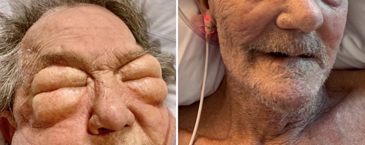
On hospital day 2, the subcutaneous emphysema had progressed and restricted the patient’s ability to open his eyes (figure 1, left). The patient began to experience obvious voice changes and upper airway stridor. CT confirmed severe bilateral subcutaneous emphysema extending into the face causing significant upper airway compression (figure 2). The patient’s oxygen saturation plummeted and endotracheal intubation was required to prevent complete airway collapse. Bilateral thoracostomy tubes were then placed to prevent worsening pneumothorax expansion and to decrease air leak into surrounding mediastinum and subcutaneous tissue. Classically, thoracostomy tube placement can potentially worsen subcutaneous emphysema and is not always recommended.4 However, this patient’s severe bullous COPD with partial right-sided lung resection allowed substantial infiltration of subcutaneous air from the pneumothorax. The patient remained mechanically ventilated for 2 days and the thoracostomy tubes remained in place for 5 days. Over the course of 7 days, the patient’s pneumothorax and compressive subcutaneous emphysema resolved. The dramatic difference is apparent in the included photos (figure 1, right), which were obtained on hospital admission and just prior to discharge.

Clinical view of facial and orbital oedema from severe subcutaneous emphysema on hospital admission (left); complete resolution of subcutaneous emphysema prior to hospital discharge (right).

CT with intravenous contrast demonstrating severe subcutaneous emphysema extending into neck and face causing significant airway compression.
Patient’s perspective
After I fell at home, I immediately knew something was wrong. I have broken ribs before, but this felt different. Within a few hours, I started to get more short of breath and when I woke up the next morning, I was swollen up like a balloon! I’m glad I went into the emergency department when I did.
The worst part about being in the hospital was having to keep the chest tubes in. I knew they were necessary to prevent any more air leaking. I am very thankful for the care I received and that all the swelling went down. Now I can breathe normal again!
Learning points
Subcutaneous emphysema rarely progresses to the point of airway obstruction.
Even with a generally benign condition such as subcutaneous emphysema, one must be cognizant of comorbidities and compounding factors that may cause serious adverse outcomes.
History and physical, along with serial examinations, are extremely important to be able to recognise acute decompensation and allow emergent management of patients with an extensive comorbid history.
Footnotes
Twitter @ctaysmith
Contributors CTS took care of patient during hospital stay, prepared photographs, wrote case outline and coordinated work of co-authors. WA took care of patient daily during admission and contributed to writing case report. MD helped finalise the case report write-up. DS assisted with obtaining background information including references.
Funding The authors have not declared a specific grant for this research from any funding agency in the public, commercial or not-for-profit sectors.
Competing interests None declared.
Patient consent for publication Obtained.
Provenance and peer review Not commissioned; externally peer reviewed.