Article Text

Statistics from Altmetric.com
Description
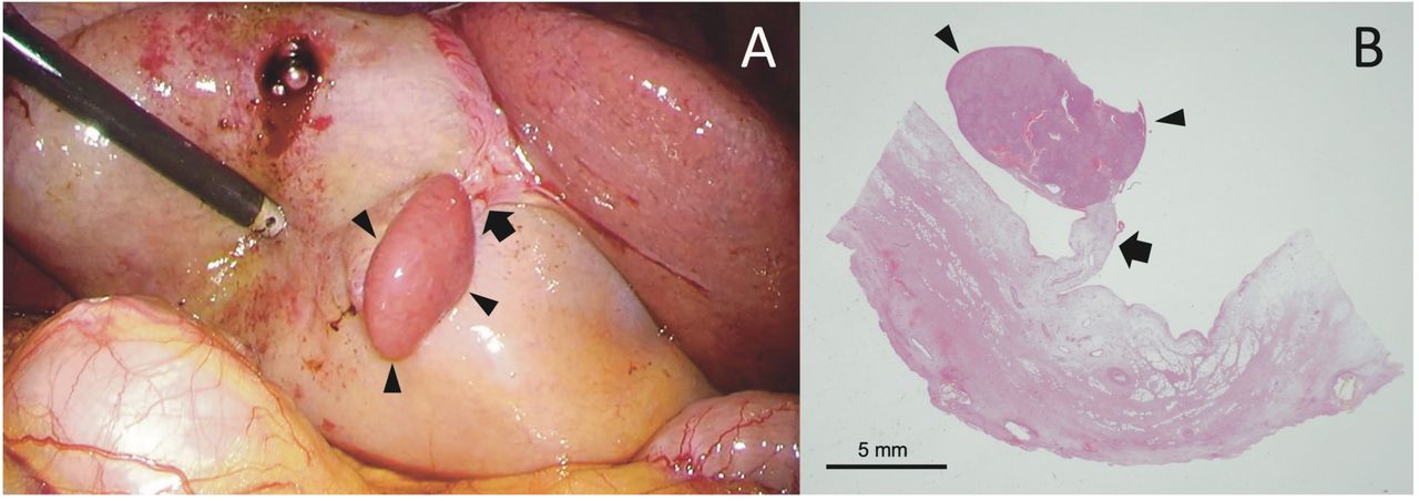
A man in his 60s was diagnosed with acute cholecystitis and referred to the surgery department for laparoscopic cholecystectomy. Operative exploration revealed an ectopic liver (EL) on the gallbladder wall (figure 1A), which was not detected by preoperative imaging. The EL stalk was dissected using an ultrasonic vessel sealer, and the EL was removed with the gallbladder. The patient was discharged with no complications, including a biliary fistula.

(A) Ectopic liver on the gallbladder wall separate from the native liver. (B) Gross observation shows a stalk containing bile ducts and blood vessels that connect the ectopic liver to the gallbladder wall. The stalk (arrow) connects the ectopic liver (arrowheads) and gallbladder wall.
EL is a rare morphological abnormality of the liver that is almost always asymptomatic and discovered incidentally during surgery or autopsy. EL is defined as liver tissue that lacks an anatomical connection to the mother liver parenchyma, often localised around the gallbladder.1 EL incidence has been reported to be from 0.24% to 0.47% at laparotomy or laparoscopy.1 Histologically, bile ducts, portal vein and arterial structures can be seen in the stalk connecting the gallbladder wall to the EL, as observed in this case (figure 1B). Here, there were no malignant findings; EL may be susceptible to hepatocellular carcinoma. Morita et al reported 39 cases of hepatocellular carcinoma in EL. They stated that the rate of cirrhosis of hepatocellular carcinoma was clearly lower than that of general hepatocarcinogenesis.2 The reason for this has been hypothesised to be related to the fact that they are less susceptible to pressure from surrounding tissues, which favours cell proliferation and incomplete vascular supply and bile excretion mechanisms.3 Resection would be desirable even on incidental detection since carcinogenesis may occur even in the absence of chronic liver disease. In addition, when resecting connective tissue around the EL, it is important to use energy devices or clips to avoid bile leakage since there may be abnormal bile duct traffic with the residual tissue.1
Patient’s perspective
I am satisfied that my illness is being adequately treated by my doctor.
Learning points
The ectopic liver is a rare morphological anomaly of the liver, and there are some reports on the relationship between the ectopic liver and hepatocellular carcinoma.
Considering the possibility of hepatocarcinoma, it is advisable to resect the ectopic liver if it is found by chance.
In addition, the stalk of the ectopic liver should be resected using energy devices or clips to address the presence of an abnormal bile duct connecting the ectopic liver to the mother liver.
Ethics statements
Patient consent for publication
Footnotes
Contributors The authors confirm their contribution to the paper: TH and KM drafted the initial manuscript and approved the final version of the manuscript. KM performed the surgery.
Funding The authors have not declared a specific grant for this research from any funding agency in the public, commercial or not-for-profit sectors.
Case reports provide a valuable learning resource for the scientific community and can indicate areas of interest for future research. They should not be used in isolation to guide treatment choices or public health policy.
Competing interests None declared.
Provenance and peer review Not commissioned; externally peer reviewed.